Cavallo Companion
Hoof Abscesses in Horses: Causes, Signs, Soaking, and Hoof Boot Support
A hoof abscess is one of the most common causes of sudden, severe lameness in horses.
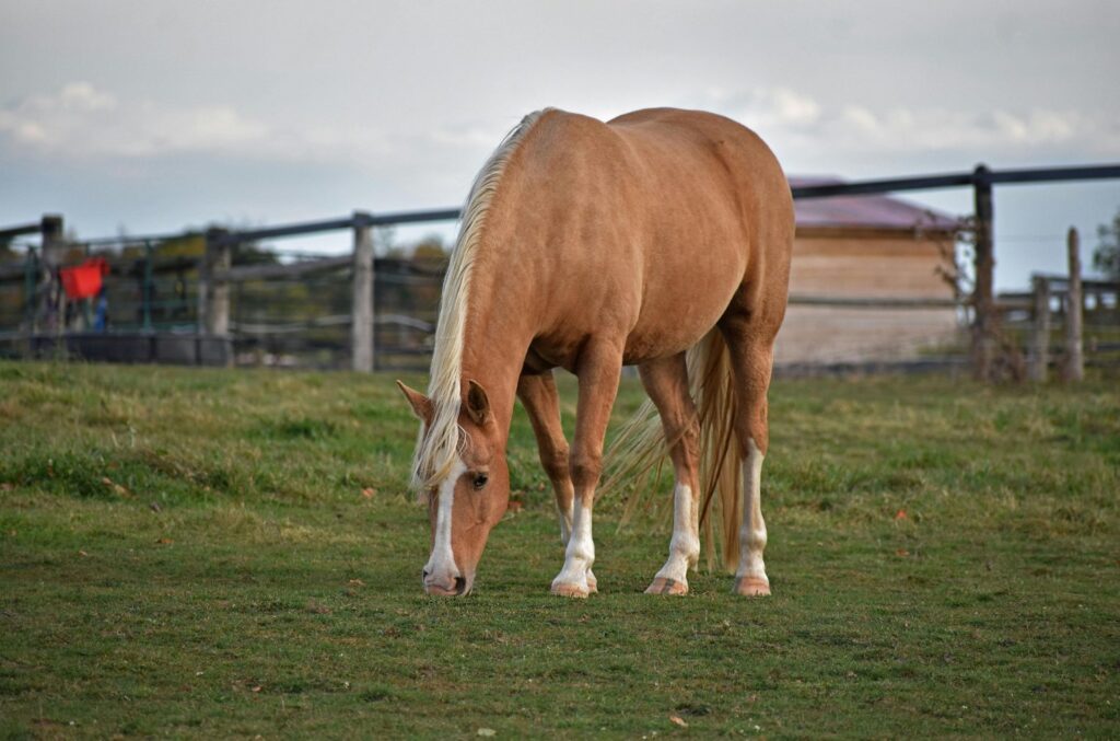
A horse may appear comfortable one day, then become reluctant to bear weight on one foot the next. This dramatic change can be alarming, and it often results from pressure building inside the hoof capsule.
Although uncomplicated hoof abscesses usually respond well once drainage is established, sudden lameness should never be diagnosed by appearance alone. Fractures, laminitis, penetrating injuries, joint infections, and soft-tissue injuries can produce similar signs. A veterinarian should assess severe or unexplained lameness and confirm whether an abscess is responsible.
What Is a Hoof Abscess?
The hoof wall, sole, frog, and bars form the outer hoof capsule. These structures consist largely of keratinized horn and surround sensitive living tissues supplied by nerves and blood vessels.
A hoof abscess develops when bacteria enter the hoof and infection forms within the sensitive tissues beneath the hoof wall or sole. The resulting inflammatory fluid, damaged cells, and infectious material collect in a confined space between the sensitive corium and the rigid hoof horn.
Unlike skin, the hoof capsule cannot expand enough to accommodate this inflammation. As fluid accumulates, pressure increases against the sensitive tissues. This explains why a relatively small abscess can produce intense pain and severe lameness.
The infection usually follows the path of least resistance. It may track beneath the sole, travel along the white line, or migrate upward through the laminar tissues. Drainage may eventually appear at the sole, white line, heel, or coronary band.
Once a drainage route opens and the pressure decreases, the horse may improve noticeably within hours. However, the drainage site still requires protection while the tissues recover and new horn develops.
How Does Bacteria Enter the Hoof?
The hoof capsule normally forms a strong physical barrier. An abscess develops when bacteria gain access through a defect in that barrier or an area of damaged tissue.
The white line is a frequent entry point. This junction connects the inner hoof wall to the sole and appears as a narrow band around the solar margin of the foot. Stretching, separation, cracks, or old nail holes can create pathways through which dirt and bacteria travel into deeper tissues.
Horseshoe nails placed inside the white line may also enter sensitive tissue and introduce bacteria. A puncture is more concerning than a straightforward abscess because it may involve deeper structures.
Bruising can contribute when repeated or concentrated pressure damages the sensitive tissue beneath the sole. Damaged tissue becomes more vulnerable to infection, particularly when bacteria enter through a nearby crack, separation, or puncture.
Factors associated with hoof abscess development include:
- White line separation or stretching
- Cracks extending through the hoof wall
- Old or misplaced horseshoe nail holes
- Direct punctures through the sole or frog
- Thin soles with limited protection over sensitive tissues
- Hoof imbalance that concentrates pressure in one area
- Bruising from hard, rocky, or uneven ground
- Repeated changes between very wet and very dry conditions
- Laminitis or other conditions that weaken white line integrity
Routine trimming supports hoof balance and limits excessive leverage on the wall and white line.
What Are the Signs of a Hoof Abscess?
The most characteristic presentation is sudden, acute lameness affecting one limb. The horse may shorten the stride, land unevenly, point the affected foot, load the toe, and avoid placing it fully on the ground or bearing weight on the affected hoof.
A stronger-than-normal digital pulse is another common finding. You can feel the palmar digital arteries along either side of the pastern. Increased blood flow and inflammation within the foot can make this pulse feel unusually strong or “bounding.” Comparing the affected limb with the opposite limb can help identify a difference.
Other possible signs include:
- Increased heat in the hoof capsule
- Pain when moving
- Sensitivity over a localized area when hoof testers are applied
- Swelling around the coronary band, pastern, or lower limb
- Drainage from the sole, white line, heel, or coronary band
- A dark tract, crack, or defect within the white line
- Reluctance to move or stand squarely
Heat is not always present, and some abscesses cannot initially be localized with hoof testers. A deep abscess may become easier to locate as it develops or moves closer to a drainage surface.
Swelling can occur when inflammation extends above the hoof capsule or when the abscess migrates toward the coronary band. However, significant swelling, fever, or illness may indicate a more extensive infection and requires prompt veterinary assessment.
Why Veterinary Diagnosis Matters
Sudden, non-weight-bearing lameness should not automatically be treated as a hoof abscess. A fracture, severe bruise, laminitis, tendon injury, joint infection, or penetrating wound may initially look very similar.
A veterinarian will usually examine the entire limb before concentrating on the foot. The assessment may include palpation, evaluation of the digital pulse, hoof-tester examination, and inspection of the sole, frog, bars, white line, hoof wall, and coronary band.
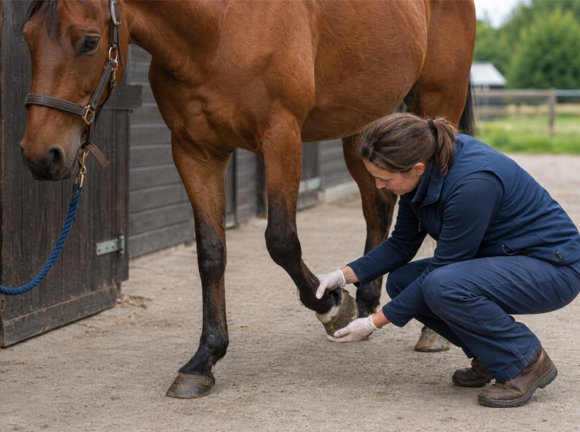
Radiographs may be recommended when the lameness cannot be localized, a penetrating injury is suspected, the horse does not improve as expected, or abscesses repeatedly occur. Imaging can help identify foreign material, gas within the tissues, reduced sole depth, changes involving the distal phalanx, or another cause of lameness.
If a nail or another object remains embedded in the hoof, leave it in place and prevent the horse from stepping farther onto it while contacting the veterinarian. The position and angle of the object provide important information about which internal structures may have been affected. Removing it before examination can eliminate valuable diagnostic evidence.
How Is a Hoof Abscess Drained?
Relieving the pressure requires a drainage route. When the tract can be accurately located, a veterinarian or qualified hoof care professional may carefully open it through the sole or white line.
They should remove only enough horn to establish drainage. Excessive hoof removal creates a larger defect, exposes more sensitive tissue, increases discomfort, and prolongs the time required for protective horn to regrow. Aggressively searching for an abscess that has not been clearly located can cause more harm than benefit.
Some abscesses are too deep to drain safely during the first examination. In these cases, warm soaking and poulticing can soften the horn and encourage the abscess to follow an existing tract toward the surface. Owners should not cut into the sole or attempt to open an abscess themselves.
In other cases, the abscess will be located, but your veterinarian or hoof care provider may opt to allow it to burst and drain naturally.
Systemic antibiotics are not automatically required for an uncomplicated hoof abscess, and your veterinarian will be able to offer personalized advice for your horse. Establishing drainage is usually the central concern because medication alone cannot relieve pressure trapped beneath the hoof capsule.
Basic Hoof Abscess Care: Soaking, Poulticing, and Protection
Warm soaking is a basic and commonly recommended part of hoof abscess care. It helps clean the hoof, soften the surrounding horn, and encourage the abscess to follow an existing tract toward the surface. Soaking can be especially valuable before the abscess opens or while the drainage tract needs to remain open.
Before soaking, pick the hoof thoroughly. Removing manure, mud, stones, and loose debris prevents this material from contaminating the soaking solution.
Then place the hoof in comfortably warm water, commonly with Epsom salts added. A typical soaking session lasts approximately 15 to 20 minutes and may be repeated 1 to 4 times daily during the active stage. The veterinarian may adjust the frequency based on the abscess location, hoof hardness and whether drainage has already been established.
A soaking boot is often easier and safer than a bucket, especially when the horse is uncomfortable or unwilling to hold the foot in one position. The boot keeps the solution around the hoof while allowing the horse to stand more naturally.
If you use a Cavallo Hoof Boot for soaking, you can temporarily cover its drainage openings with waterproof tape before adding the soaking solution. Another option is to place a sturdy plastic bag over the hoof, followed by a hoof boot, then fill the bag with warm water once it has been secured. The horse should remain supervised throughout the soak, and the boot should be removed immediately afterward. Clean and dry the hoof and boot.
Never leave a sealed boot containing liquid on for turnout or extended wear.
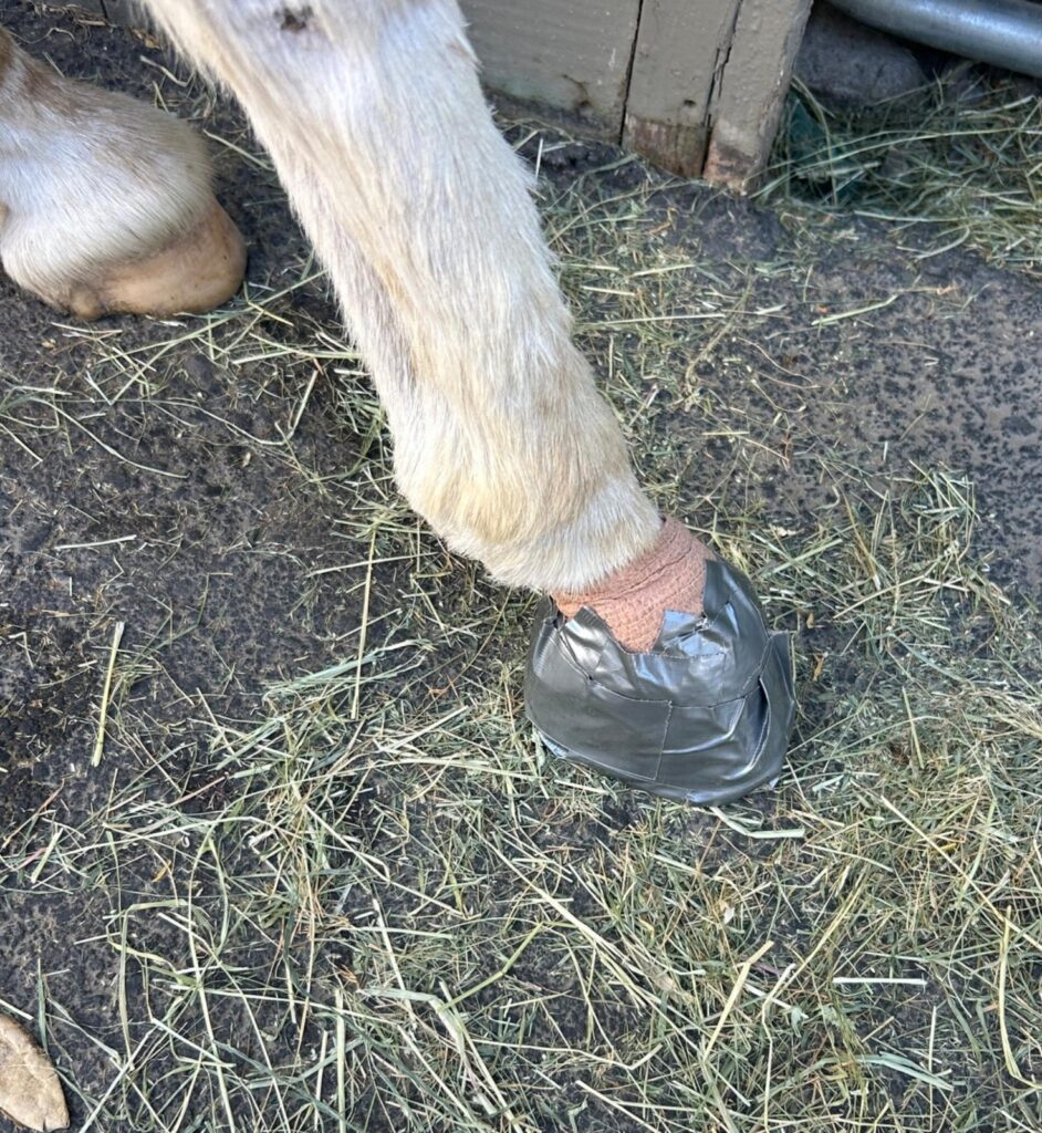
After soaking, a wet or drawing poultice may be applied to help maintain a moist environment and support continued drainage. Secure the poultice without restricting circulation or placing excessive pressure around the coronary band and pastern. A common method is using a diaper and/or vet wrap to cover the hoof, followed by a diy duct tape cover, or an easier option, such as a hoof boot.
Once the drainage and discharge have decreased, care usually moves from drawing moisture outward to keeping the area clean and protected. A dry dressing can help prevent mud, manure, stones, and contaminated bedding from entering the opening while the sole begins to close.
Soaking should continue for a clear purpose, not indefinitely. Prolonged exposure to moisture after drainage is complete can soften the sole, frog, and hoof wall. Veterinary guidance can help determine when soaking is no longer needed and clean, dry protection becomes more appropriate.
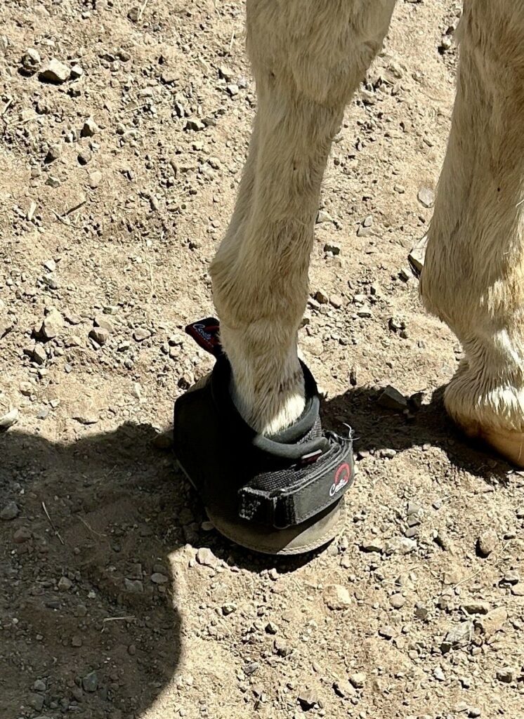
Want to hear from people who use Cavallo hoof boots for abscesses? This is what Larry from California has to say: “I received the D-1 Slim LEBs. Thank you for getting them out and to us so quickly! The boots are to help recover from an abscess. They sure beat messing around with vet wrap and duct tape! Thought I would share a couple photos of what we previously had to do and then with him wearing a boot. So far, so good 🤞”
Keeping the Horse Comfortable During Recovery
A horse with a hoof abscess should be kept on clean, supportive footing. Deep bedding can reduce pressure on the painful foot and make it easier for the horse to lie down. Food and water should remain within easy reach when the horse is reluctant to walk.
Do not force the horse to move to encourage drainage. Controlled movement may become appropriate as comfort improves, but this depends on the location of the drainage opening, the condition of the sole, and the veterinarian’s recommendations.
Once the abscess has opened, change the dressing whenever it becomes wet, displaced, or soiled. Each dressing change provides an opportunity to examine the hoof and assess:
- The amount and appearance of drainage
- Hoof temperature
- The strength of the digital pulse
- Swelling around the coronary band or lower limb
- The horse’s willingness to bear weight
- The condition of the drainage opening
- Any unusual or persistent odour
Pain medication may be prescribed when necessary. It should be given under veterinary direction because improved comfort does not always mean the infection has resolved or the drainage site is ready for unrestricted movement.
How Do Hoof Boots Support Abscess Recovery?
A hoof boot does not remove the infection or drain the abscess. Its role is to protect the tender solar surface and drainage opening while the hoof recovers.
Before drainage, pressure within the hoof can make contact with firm ground extremely painful. After drainage, the opening and surrounding sole may remain sensitive until protective horn develops. A properly fitted hoof boot creates a barrier between the foot and hard, rocky, frozen, or uneven footing.
A boot can also hold a poultice, dressing, or protective pad in position. A suitable pad may distribute load across a broader area of the sole and reduce concentrated pressure around the drainage site. The pad should not press directly into the opening, and its thickness should not make the boot tight or unstable.
When using a hoof boot during abscess recovery:
- Allow enough room for the dressing without compressing the hoof, heel bulbs, or pastern.
- Remove the boot at least daily and more frequently while drainage is active.
- Replace wet or contaminated dressings and pads promptly.
- Check the heel bulbs, pastern, sole, and coronary band for rubbing, swelling, heat, or trapped moisture.
- Clean and dry the boot before replacing it.
- Reassess the fit whenever the dressing or pad thickness changes.
If a thick dressing or pad raises the affected foot, a matching boot and pad may be appropriate on the opposite hoof. This can reduce the height difference between the two limbs, particularly when boots will be worn for an extended period. A veterinarian or hoof care provider can determine whether this is necessary.
Cavallo Hoof Boots can serve two practical purposes during this process. They can be temporarily adapted for soaking and then used to protect the dressed hoof afterward. Always clean and dry the boot between uses.
Dr. Robert Judd is a veterinarian based in Hewitt, Texas. He recommends Cavallo boots to his clients when treating hoof abscesses, as an easy alternative to time-consuming and expensive wrapping supplies.
“I purchased a quarter horse/Friesian cross as a trail horse that was basically a rescue. The horse has severe thrush in all 4 feet on the solar frog and the frog at the central sulcus. This horse needed his feet treated daily for 2 weeks and then every other day. To avoid using so much wrap material and spending the extra time to wrap, I purchased the ELB boots for all 4 feet. These boots worked great and are durable. They have a hole in the side so the horse does not sweat in the Texas heat like some boots. I simply treated the foot and applied a small wrap over the sole and applied the boot. The horse wore the boots 24/7 for 30 days with no issues. I think using these boots for treating a hoof problem is a good investment and saves time and money. I will recommend these boots to my clients for treating solar abscesses and other foot problems rather than extensive wrapping.”
When Does an Abscess Need Further Investigation?
Many uncomplicated hoof abscesses become considerably less painful after adequate drainage. If the expected improvement does not occur, reconsider the diagnosis or drainage route.
Veterinary reassessment is important when:
- The horse remains unwilling to bear weight after drainage.
- Lameness improves and then returns.
- Swelling progresses above the hoof.
- The horse develops a fever or appears unwell.
- Drainage continues, smells unusually foul, or repeatedly returns.
- A nail or another penetrating object may have entered the hoof.
- More than one hoof becomes painful.
- The horse has a history of laminitis or metabolic disease.
- Abscesses occur repeatedly.
Persistent drainage may mean the tract closed before the abscess emptied completely. It can also indicate retained foreign material, a deeper infection, white line disease, a keratoma, or involvement of the distal phalanx.
Repeated abscesses deserve a broader assessment of the hoof and the horse’s health. The veterinarian and hoof care provider may examine sole depth, white line integrity, hoof balance, wall cracks, old nail holes, laminar health, and areas of concentrated pressure. Turnout conditions, trimming practices, nutrition, and repeated wet-to-dry environmental changes should also be reviewed.
Regular, balanced trimming; preserving functional sole; prompt attention to cracks and white line separation; and appropriate hoof protection can reduce avoidable risks. However, even well-managed horses can occasionally develop an abscess following a bruise, puncture, or small defect in the hoof capsule.
Supporting a Horse Through a Hoof Abscess
A hoof abscess becomes painful because infection and inflammatory material are trapped inside the rigid hoof capsule. Once the abscess has been accurately identified, basic care focuses on warm Epsom salt soaking, establishing and maintaining drainage, applying a clean poultice or dressing, and protecting the tender foot from pressure and contamination.
A properly fitted Cavallo Hoof Boot can make soaking easier, hold a dressing securely, and protect the sole as the horse becomes comfortable enough to move normally. It provides practical support during recovery, but it does not replace an accurate diagnosis or veterinary care.
Most uncomplicated abscesses improve considerably after the pressure has been relieved. Continued pain, progressive swelling, recurring drainage, or repeated abscesses indicate that the foot requires further veterinary investigation.
Ready to choose?
Find the Right Boot for Your Horse
A properly fitted hoof boot protects the tender sole and the drainage opening, and holds a dressing in place while the hoof recovers. Measure the hoof, then choose the boot that suits your horse.
Sources
- Redding, W. R., and A. E. O’Grady. “Septic Diseases Associated with the Hoof Complex: Abscesses, Punctures, and Keratomas.” Veterinary Clinics of North America: Equine Practice, vol. 28, no. 2, 2012, pp. 423–440.
- Turner, T. A. “Diagnosis and Treatment of the Equine Foot.” Veterinary Clinics of North America: Equine Practice, vol. 19, no. 2, 2003, pp. 309–332.
- Cuomo, F. “Horse Hoof Abscesses.” University of Minnesota Extension, reviewed 2021.
- American Association of Equine Practitioners. “Front Foot Lameness: Don’t Forget the Exam.” AAEP Horse Health Resources.